


 微信公众号
微信公众号

News Center
Latest Guidelines from the National Health Commission: Hereditary Factors, Classification, and Clinical Manifestations of Kidney Cancer
May 18,2022
Renal cell carcinoma (RCC) is a malignant tumor originating from the renal tubular epithelium, accounting for 80% to 90% of malignant tumors of the kidney. The most common histopathological type of renal cell carcinoma is clear cell carcinoma, followed by papillary renal cell carcinoma and chromophobe carcinoma, as well as rare types such as collecting duct carcinoma. With the development of medical imaging, the detection rate of early renal cell carcinoma has gradually increased. Localized renal cell carcinoma can achieve satisfactory efficacy through nephron-sparing tumor resection or radical nephrectomy (RN).
According to the GLOBOCAN 2020 global cancer statistics, in 2020, the incidence of renal cell carcinoma ranked 14th among malignant tumors worldwide, lower than prostate cancer and bladder cancer in the urinary system, and the mortality rate ranked 15th.
Recently, the "Guidelines for the Diagnosis and Treatment of Renal Cell Carcinoma (2022 Edition)" were released. This article summarizes parts of the etiology and clinical manifestations of renal cell carcinoma.
Etiology of renal cell carcinoma
Not yet clear; the occurrence is related to genetics, smoking, obesity, and other factors.
1. Genetic factors
Most renal cell carcinomas are sporadic; hereditary renal cell carcinoma accounts for 2% to 4% of all cases, mostly inherited in an autosomal dominant manner within families, caused by different genetic mutations involving both tumor suppressor genes and oncogenes. Known hereditary renal cell carcinomas include von Hippel-Lindau (VHL) disease (bilateral multiple clear cell renal carcinomas and renal cysts), MET gene-related hereditary papillary renal cell carcinoma, hereditary leiomyomatosis and renal cell carcinoma caused by fumarate hydratase gene abnormalities, Birt-Hogg-Dubé (BHD) syndrome (multiple chromophobe renal cell carcinomas, hybrid chromophobe and eosinophilic renal tumors, papillary renal cell carcinoma), and HRPT2 gene-related hyperparathyroidism-jaw tumor syndrome (mixed epithelial and stromal tumors, papillary renal cell carcinoma) (Table 1).
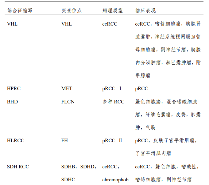
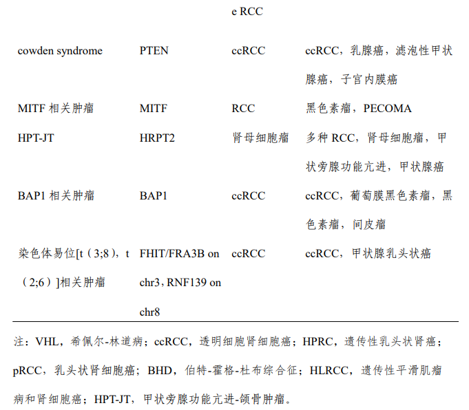
The following groups are generally considered potential patients with hereditary renal cell carcinoma:
① Patients with renal cell carcinoma aged ≤45 years;
② Bilateral/multiple renal tumors;
③ Family history of renal cell carcinoma (at least one first-degree relative, at least two second-degree relatives);
④ Renal cell carcinoma combined with other tumor histories (pheochromocytoma, gastrointestinal stromal tumor, central nervous system hemangioblastoma, pancreatic neuroendocrine tumor, etc.), combined with other lesions such as lung cysts, spontaneous pneumothorax, etc.;
⑤ Combined with rare skin lesions (leiomyoma, angiofibroma, etc.);
⑥ Personal or family history of renal cell carcinoma-related syndromes. For these patients, genetic mutation testing for themselves and related family members can be recommended.
2. Smoking
Smoking can increase the risk of renal cell carcinoma. Prospective studies consider smoking a moderate risk factor. The relative risk of renal cell carcinoma is 1.3 for former smokers and 1.6 for current smokers.
3. Obesity
Obesity is generally expressed by body mass index (BMI); as BMI increases, the risk of renal cell carcinoma increases. The specific mechanism by which obesity increases the risk of renal cell carcinoma is unclear but may be related to increased release of androgens and estrogens or cytokines released by adipocytes.
4. Acquired renal cysts related to long-term dialysis in end-stage renal disease
Compared with the general population, patients with end-stage renal disease have a higher incidence of renal cell carcinoma. Patients on long-term dialysis are prone to acquired renal cysts. In these renal cell carcinoma patients, tumors are usually bilateral and multiple, with papillary histological structures.
Other evidence suggests that alcohol consumption, occupational exposure to trichloroethylene, and women with high estrogen levels may also increase the risk of renal cell carcinoma. Further research is needed on the potential interaction between genetic factors and environmental exposures.
Clinical manifestations of renal cell carcinoma
The clinical manifestations of renal cell carcinoma patients are complex and variable. Some of these manifestations are directly caused by the renal tumor itself, while others may be due to hormones secreted by cancer cells or metastatic lesions. With the increasing popularity of health check-ups, most renal cell carcinoma patients visiting hospitals are usually incidentally discovered by imaging examinations.
In clinical practice, early renal cell carcinoma often lacks clinical manifestations. When the classic triad of renal cell carcinoma (hematuria, flank pain, and abdominal mass) appears, most patients are already in the middle or late stages; the presence of left-sided varicocele suggests possible left renal vein tumor thrombus; therefore, early diagnosis of renal cell carcinoma is of great significance.
>>>> Paraneoplastic syndrome
Clinical manifestations are not directly caused by the primary tumor or metastatic site but are due to abnormal immune responses induced by tumor-secreted products or other unknown reasons, causing pathological changes in endocrine, nervous, digestive, hematopoietic, musculoskeletal, renal, and skin systems, with corresponding clinical manifestations, known as paraneoplastic syndrome. The incidence of paraneoplastic syndrome in renal cell carcinoma patients is about 30%, presenting as hypertension, elevated erythrocyte sedimentation rate, polycythemia, liver dysfunction, hypercalcemia, hyperglycemia, neuromuscular disorders, amyloidosis, galactorrhea, coagulation abnormalities, etc. Patients with paraneoplastic syndrome have a poorer prognosis.
>>>> Symptoms caused by metastatic lesions
Some patients with renal cell carcinoma present initially with clinical manifestations of metastatic lesions, such as bone pain, fractures, cough, hemoptysis, etc. Physical examination may reveal cervical lymphadenopathy, secondary varicocele, and bilateral lower limb edema, the latter suggesting possible tumor invasion of the renal vein and inferior vena cava. Among patients with metastatic renal cell carcinoma, the common metastatic organs and their incidence rates are as follows: lung metastasis (48.4%), bone metastasis (23.2%), liver metastasis (12.9%), adrenal metastasis (5.2%), skin metastasis (1.9%), brain metastasis (1.3%), and other sites (7.1%). Advanced patients may also exhibit cachexia symptoms such as weight loss, fatigue, and poor appetite.
Source: Yimai Oncology Department
Previous:
Contact Us
Consultation phone: 400 000 8876
Recruitment service: pingping@calt-bio.com
Business cooperation: pingping@calt-bio.com
Company address: Building A8, Huizhi Technology Park, High-tech Industrial Park, Nanjing